Or , our new artificial intelligence tool.
MyEni Login
Experimental geosciences: digital microscopes and technologies
Light and electron microscope images analysed and integrated with digital technologies reduce the time for rock characterisation.

What are new experimental geosciences?
The development of innovative technologies capable of meeting the challenges posed by increasingly complex assets is essential when it comes to studying the subsurface. We apply the digital methodologies of pattern recognition and machine learning techniques to optical analysis. In the field of electron microscopy, we have developed the PICADOR (Porosity Image Analysis of Reservoir Rocks), a proprietary approach focusing on the characterisation of the porous system, i.e. the set of microscopic pores in reservoir rocks that, a bit like a 'sponge', retain natural gas and other hydrocarbons within them. Using the PICADOR system, in particular, we can reconstruct the geometric characteristics of porous systems through images taken from thin rock sections. This approach can also be used in the context of CO₂ capture and storage (CCS) to quantify the effects of carbon dioxide injection.
A further cognitive approach we have developed is the MINSYS (Mineralogy Site Specific), focusing on determining the chemical compositions of site-specific minerals.
In this case, the information gathered makes it possible to obtain the thermodynamic parameters of the ores themselves, which are then used for the numerical modelling of flow and transportation and transportation of fluids. This modelling takes into account the chemical and physical interactions between rock and fluid, is performed with the MuFloTSw (Multiphase Flow and Transport in Homogeneous and Fractured Systems) proprietary software.


Industrialisation
Carbon management
Geophysical exploration
Natural gas and other hydrocarbons
New analytical technologies
The use of digital technologies has made the analysis process more efficient and versatile, allowing us to study even smaller sample sizes. Our laboratories are equipped with a triaxial device, one of only three in the world, which is capable of performing full geomechanical analyses on 1 cm rock samples.
Other fundamental analysis tools for small samples are the X ray microtomography and digital rock physics. These make it possible to obtain a static three-dimensional image of the porous space of the rock under study and to conduct virtual laboratory experiments. The result is similar to that obtained by the experiment on a real rock sample, but with extremely reduced execution time and material consumption. All this information is complementary and is supplemented with images collected by the electron microscope (SEM).
-
Sample analysed by the X-ray microtomography
-
Tomography for the 3D view of the movement of fluids inside the rock samples
-
Computerised Axial Tomography (CAT) of cores to characterise the rock of the subsoil
-
Rock sample
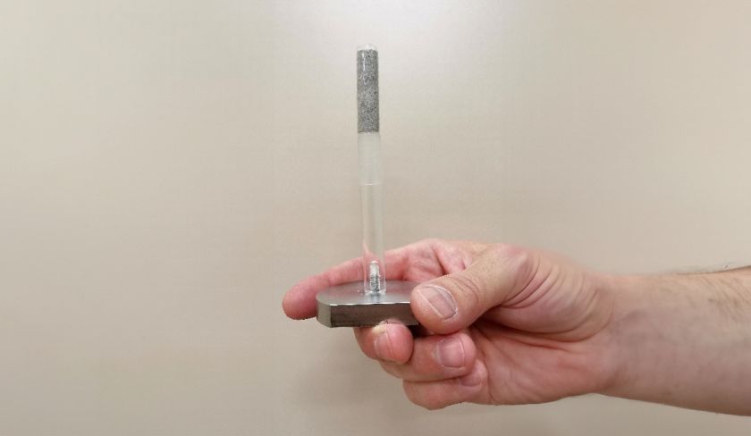
Sample analysed by the X-ray microtomography
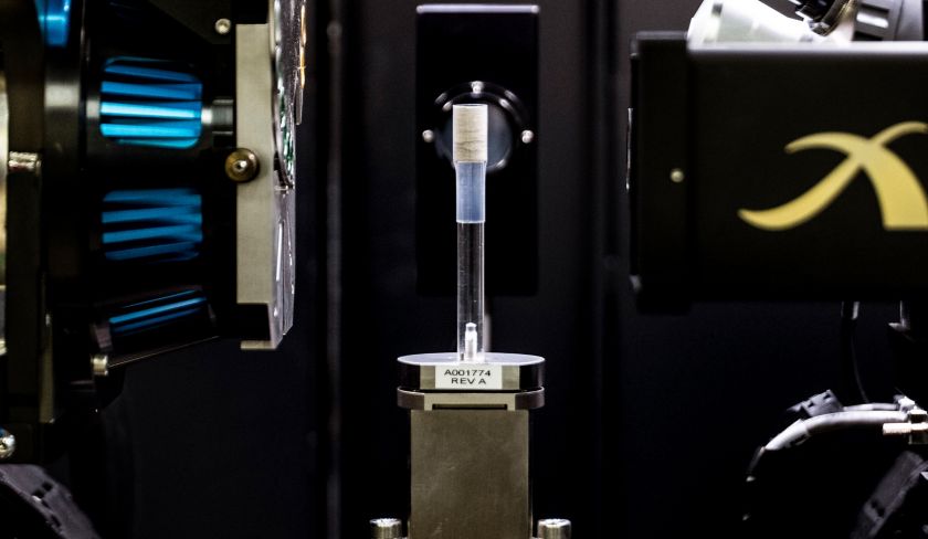
Tomography for the 3D view of the movement of fluids inside the rock samples
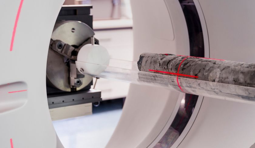
Computerised Axial Tomography (CAT) of cores to characterise the rock of the subsoil
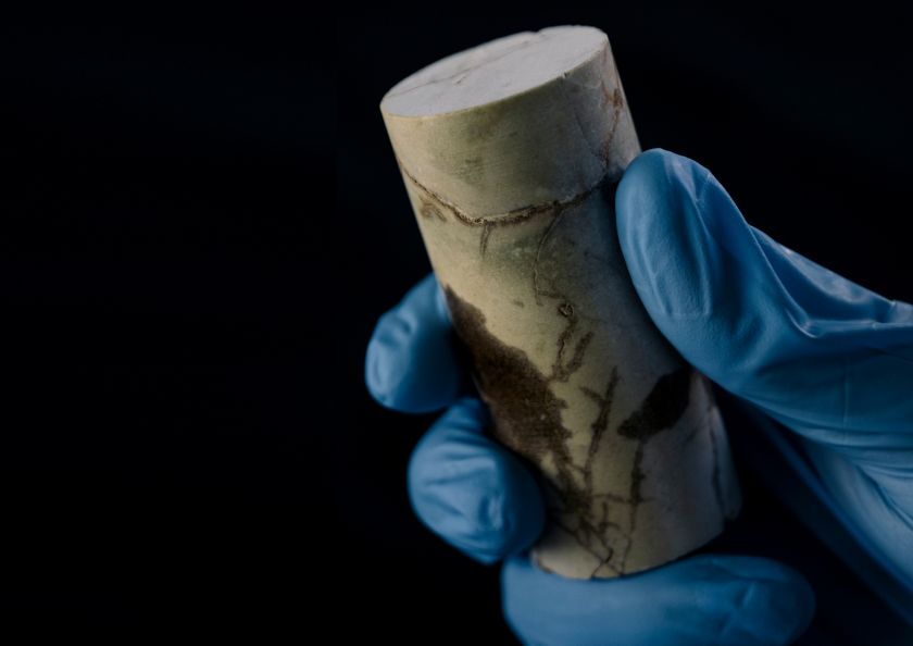
Rock sample
Our laboratories are also equipped with proprietary software, including the 4D-CoreINV©, which allows us to interpret dynamic experiments performed under a 3D tomography and instantly extract much of the input data required for numerical simulation.
Unlike the experiments conducted with the microtomography, these are dynamic because they consist of injecting fluids into the rock sample and filming its movement in 3D with a resolution of 50 microns. Thanks to Eni's HPC supercomputer, the software processes the amount of data acquired (approx. 10 million numbers) in just a few hours.
Features and performance
-
~4.000metres deep
of coring wells
-
>1000analyses
carried out per core sample
-
~1.5inches
sample sizes used for analysis
-
50microns
resolution of geomechanical analysis of the triaxial device
How we sample the rock
Our analyses are based on rock samples extracted through the coring, which consists of digging small yet very deep exploratory well, from which so-called 'cores', i.e. long cylindrical sections of underground rock, are extracted. From cores taken from wells at a depth of around 4,000 metres and brought to the surface, it is possible to extract a wealth of invaluable information, using both experimental and digital protocols. We can run up to 1,000 analyses per core sample, obtaining a detailed characterisation of the pores, their interconnections and fluid interactions. In our core archive we have stored hundreds of kilometres of core samples.
To limit costs and operational risks and promote the sustainability of our activities, we are now able to extract small cores from the walls of wells (approximately 1-1.5 inches). These cores, properly sampled thanks to new technologies, instrumentation, analytical protocols, and constantly updated skills, allow us to run the same analyses and characterisations as those carried out on traditional cores.
